 |
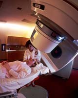
Washington (UPI) Dec 22, 2006 Elastography, a non-invasive ultrasound imaging method that can distinguish between benign and malignant tissues, is now on the U.S. market. The new technique uses a regular ultrasound apparatus with special software attached. Tissue being explored is manually compressed very lightly, and the tissue's response to this compression -- or its elasticity -- is measured using ultrasound echoes and made into a picture called an elastogram. "The important hypothesis here is that tissue elasticity is related to disease," Jonathan Ophir, the developer of this technique, told United Press International. The light areas on the elastogram represent softer tissue, with the dark areas indicating stiffer structures. The elastogram and sonogram can be displayed side-by-side on a screen and cursors can be moved simultaneously in both images when an area is being evaluated. Ophir's laboratory is located at the University of Texas Medical School in Houston, where he and his colleagues have been working on the elastography for over 15 years in collaboration with other laboratories worldwide. "Pathological tissues, especially malignant tumors, are usually harder than normal tissue because their quickly proliferating cells make them more dense. In addition, malignant tumors of the breast appear to develop areas of stiff scar tissue around them. Elastograms pick up this scarred area, but regular ultrasound images do not." Ophir said that exploring why elastograms consistently produced images of breast tumors that were larger than those created by regular ultrasound techniques led researchers to realize the device's diagnostic potential for breast cancer. Elastography was picking up the scarred area around malignancies, and this proved to be a promising way to differentiate a benign from a cancerous tumor in the breast. "Brian Garra at the University of Vermont has done all of our clinical work," Ophir said. "He started working on breast disease because currently, every palpable lump is biopsied and 80 to 90 percent of them turn out to be benign. That's a lot of unnecessary pain and suffering and it costs between one and two billion dollars per year in the U.S. alone." Since Garra's work was published in 1997, Richard Barr, a Youngstown, Ohio-based researcher has tested the technique on about 100 breast-disease patients whose tumors were later biopsied to confirm the diagnosis. Elastograms correctly identified nearly 100 percent of the malignancies present based on size difference alone, he noted. Similar results were obtained earlier in England by William Svenssen at the Charing Cross Hospital in London. Ophir said that, while manual palpation is an age-old method of finding stiffer tissue, it is limited to structures that are close to the skin and large enough to be felt. Elastography allows physicians to find deep structures as small as several millimeters in diameter. It does not work on lungs or other tissues surrounded by bone that does not allow compression, researchers said, but may be useful for exploring muscles, thyroid, and breast and prostate tissues. Ophir said he and his colleagues have been working on the technique since 1989, when a series of failed experiments prompted them to explore other options. "(The National Institutes of Health) gave us a grant in the late 1980s to develop a way to measure the speed of sound in tissues," Ophir told UPI. "We found that the speed of sound doesn't change much from tissue to tissue and the elasticity of the tissues produced a great deal of error in our measurements. "One morning we woke up and said 'Why don't we make a picture of tissue elasticity instead of the speed of sound?'" Their first picture was produced in 1991. "It was very primitive," Ophir said. "Ultrasound equipment wasn't sophisticated back then. We were using home-brew instruments, but we were able to show that it was possible." They coined the term "elastography" in 1991, he added, when they realized the images they were producing were no longer sonograms, but a unique new tool. Since then, Ophir and his colleagues have taken advantage of every advance in ultrasound and computer technology to improve their device. "The acoustical and mechanical properties of the tissue, how you process the ultrasound signals, and how much compression must be applied to obtain quality images are all important," Ophir said. "There are a dozen or more different parameters that influence the final image quality." He said obtaining the image was also a trial-and-error process. The team previously used a mechanical device to get the 1-percent compression that produces the best picture, but several years ago, researcher Jeff Bamber and his co-workers at the Institute of Cancer Research in Sutton, England, showed that handheld devices worked as well when the operator watched the image on the screen and adjusted the pressure accordingly. The new technique has recently gone public. Siemens introduced an instrument based on its Antares ultrasound machine this November at the annual meeting of the Radiological Society of North America in Chicago, Ill. Hitachi Medical, based in Tokyo has developed an elastography device that is marketed in Europe and Japan, and Medison of Korea has also developed an instrument. Ophir said he anticipated that other major players will soon follow suit as well. He said he expects the cost to be under $100,000, noting that that price tag is considered mid-range for ultrasound equipment. More information on elastography can be accessed at the official University of Texas Medical School elastography Web site at www.elastography.com. Community Email This Article Comment On This Article Related Links Hospital and Medical News at InternDaily.com Nuclear Space Technology at Space-Travel.com
 Dallas (UPI) Dec 28, 2006
 More than one of every three Americans die with cardiovascular disease as the underlying cause, according to new data form the American Heart Association (AHA). |
|
| The content herein, unless otherwise known to be public domain, are Copyright 1995-2006 - SpaceDaily.AFP and UPI Wire Stories are copyright Agence France-Presse and United Press International. ESA PortalReports are copyright European Space Agency. All NASA sourced material is public domain. Additionalcopyrights may apply in whole or part to other bona fide parties. Advertising does not imply endorsement,agreement or approval of any opinions, statements or information provided by SpaceDaily on any Web page published or hosted by SpaceDaily. Privacy Statement |