 |
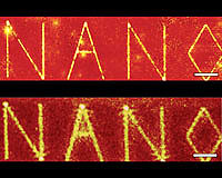
Austin TX (SPX) Sep 20, 2006 A microscope used to scan nanostructures can be dramatically enhanced by using a 'superlens,' reports an international team of scientists from the Max Planck Institute (MPI) for Biochemistry and The University of Texas at Austin in this week's issue of Science. This is the first time a superlens, a lens capable of creating images of objects smaller than the wavelength of light, has been integrated into a microscope and used to visualize two-dimensional objects. The team of scientists produced images of holes in a gold film that were smaller than a wavelength of light, about 500 nanometers in diameter, by equipping a near-field scanning optical microscope with a newly fabricated superlens. "Though the idea of a superlens was invented in 2000, it has been unclear how, or if, a superlens could be integrated into an imaging system," says Gennady Shvets, associate professor of physics at The University of Texas at Austin. "Nor has it been demonstrated that ultra-small two-dimensional objects, such as holes or nanodiscs, can be imaged by a superlens. "We were able to image a two-dimensional object--holes in a surface. Such imaging would be impossible without the superlens." The resolution of conventional optical microscopes is limited to about half the wavelength of the light used to illuminate an object. The superlens was invented by Sir John Pendry in 2000 to overcome that limit and make images of very tiny objects. The new superlens was fabricated at The University of Texas at Austin using specialized equipment at the Center for Nano and Molecular Science and Technology and the J.J. Pickle research campus. The lens was made of a thin film of silicon carbide and was sandwiched between two layers of silicon oxide. A layer of gold only 60 nanometers thick pocked with holes of various sizes was attached to the bottom of the lens. Researchers Thomas Taubner and Rainer Hillenbrand at MPI then used the Texas superlens with a near-field scanning optical microscope to create an image of the holes in the gold layer beneath the surface of the lens. Near-field scanning optical microscopy (NSOM) works by passing a tiny probe across the surface of a specimen, producing an image of the surface line by line. While the resolution attained by NSOM is very good, it can record only the surface of a nanoscale object. The action of the scanning probe in contact with the surface also makes it difficult to visualize fragile specimens, such as living cells. Adding the superlens to NSOM not only increases the resolution of the image, but also provides a barrier between the scanning probe and the object. "This is the first attempt to integrate a superlens into a scanning optical microscope, something that could be very useful in industrial applications," says Shvets. "One new possible application could be the imaging of biological objects in their natural environment, separated by a superlens from the probing tip." Shvets says another application of the superlens could be the non-destructive probing of metallic interconnects buried under a layer of glass, as is commonly used in semiconductor devices. Shvets cautions there is much work to be done before applying the technique widely. His group at The University of Texas at Austin aims to make thinner silicon carbide films (50-100 nanometers thick) that will provide even higher resolution images. Other authors of the paper from The University of Texas at Austin are Dimitriy Korobkin, a research scientist in the Physics Department, and Yaroslav Urzhumov, a physics graduate student. Community Email This Article Comment On This Article Related Links Physics at UT Nano Technology News From SpaceMart.com Nano Technology News From SpaceMart.com Computer Chip Architecture, Technology and Manufacture
 Baltimore MD (SPX) Sep 20, 2006
Johns Hopkins researchers have devised a way to use a brief burst of electricity to release biomolecules and nanoparticles from a tiny gold launch pad. The technique could someday be used to dispense small amounts of medicine on command from a chip implanted in the body. The method also may be useful in chemical reactions that require the controlled release of extremely small quantities of a material. |
|
| The content herein, unless otherwise known to be public domain, are Copyright 1995-2006 - SpaceDaily.AFP and UPI Wire Stories are copyright Agence France-Presse and United Press International. ESA PortalReports are copyright European Space Agency. All NASA sourced material is public domain. Additionalcopyrights may apply in whole or part to other bona fide parties. Advertising does not imply endorsement,agreement or approval of any opinions, statements or information provided by SpaceDaily on any Web page published or hosted by SpaceDaily. Privacy Statement |