 |
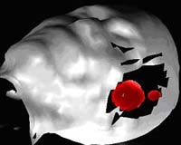
Pasadena (JPL) Jul 15, 2004 Using an infrared video camera developed by researchers at NASA's Jet Propulsion Laboratory in Pasadena, Calif., surgeons are testing thermal imaging and image processing to see if they can create useful maps of brain tumors. Researchers want to see if the camera, which detects infrared -- or heat -- emissions might help neurosurgeons better visualize tumors before they operate and also find tiny clusters of cancerous cells that might remain after surgery. NASA scientists already use infrared technology to map Earth's surface and search for distant objects in the universe. Firefighters use it to locate people trapped in buildings, and military forces track down their targets hiding in the dark. Physicians have used infrared technology for mapping the roots of skin cancer, but it has never been used for brain tumors until now. Doctors at the Keck School of Medicine of the University of Southern California in Los Angeles are using the JPL- developed camera and infrared imaging in a trial. They're trying to see if they can sketch tumor margins by detecting temperature changes during surgery, since tumor cells emit more heat than healthy ones. "The camera's precision allows it to map temperature differences of one-hundredth of a degree Celsius at a high resolution," said Dr. Sarath Gunapala, JPL lead engineer for the camera. Currently, neurosurgeons delve carefully into the brain and remove as much of the tumor as they can see under magnification. However, they may take healthy tissue along with the cancerous cells or leave residual cells that can grow back along the tumor's margins. "Brain tumor tissue looks the same as healthy tissue on the edges," said Babak Kateb of the Keck School of Medicine, a research fellow and lead scientist of the project. "Tumor cells use different biochemical pathways from normal cells, and when researchers use the infrared camera, they can pick up hotspots or areas of tissue warmer than normal tissue." After doctors receive infrared images of the brain, image processing software marks the boundaries between tumor regions and surrounding healthy tissue. "We are refining software similar to what our group has been using for analyzing rocks on Mars and other planets," said Dr. Wolfgang Fink, JPL senior researcher. "An advantage of thermal imaging is that it's non-invasive," said Dr. Peter Gruen, a neurological surgeon at the Keck School of Medicine. "It measures heat energy emerging from patients without exposing them to X-rays or intravenous solutions, and is performed without incisions or contact to the brain tissue." Community Email This Article Comment On This Article Related Links Keck School of Medicine SpaceDaily Search SpaceDaily Subscribe To SpaceDaily Express Space Medicine Technology and Systems
 Richland WA (SPX) Jun 22, 2005
A new computational tool developed at the Department of Energy's Pacific Northwest National Laboratory is speeding up our understanding of the machinery of life - bringing us one step closer to curing diseases, finding safer ways to clean the environment and protecting the country against biological threats. |
|
| The content herein, unless otherwise known to be public domain, are Copyright 1995-2006 - SpaceDaily.AFP and UPI Wire Stories are copyright Agence France-Presse and United Press International. ESA PortalReports are copyright European Space Agency. All NASA sourced material is public domain. Additionalcopyrights may apply in whole or part to other bona fide parties. Advertising does not imply endorsement,agreement or approval of any opinions, statements or information provided by SpaceDaily on any Web page published or hosted by SpaceDaily. Privacy Statement |