 |
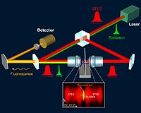
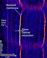
Munich - Apr 11, 2002 Max Planck researchers demonstrate a 15-fold increase in axial resolution in fluorescence 3-D light microscopy - the first breakthrough of optical focusing microscopy into the nanometer range At some point in physics class, we all learn that a light microscope cannot display details smaller than the wavelength of light. Objects closer to each other than about a quarter of a micrometer will therefore blur - no matter how perfect the microscope. In the late 19th century, physicist Ernst Abbe found an explanation for this phenomenon: light is a wave and therefore diffraction does not permit us to focus light onto an area smaller than on third of the wavelength of the light. The basic nature of this problem led to the development of the electron microscope, and, more recently, of the scanning tunneling microscope as well as the atomic force microscope. Today, these microscopes have an extremely high performance and a high enough resolution to display features on a molecular level. Such microscopes tend have one crucial drawback however, insofar as they are usually limited to viewing the surface of a specimen or to viewing a very thin layer. It is due to this fact that light microscopes have remained indispensable in biological and medical research. Not only do they provide three-dimensional images of live cells, they can also map out biochemical processes inside the cell. For this reason, Stefan Hell's High Resolution Microscopy research group at the Max Planck Institute for Biophysical Chemistry in G�ttingen, Germany, pursued the challenging goal of overcoming these century-old limitations - namely in the area of fluorescence. Fluorescence microscopy permits us to target as well as to mark specific sub-cellular components with dyes. After exposing them to light, they fluoresce, thereby disclosing their exact location to us. Recently, the research team demonstrated that it is possible to narrow the focal spot of the fluorescence microscope by applying stimulated emission, a phenomenon well known in laser physics. Immediately after being excited by a (green) light pulse, they are de-excited by a subsequent stimulating (red) light pulse (Stimulated emission deletion, STED). The de-exciting pulse is aligned around the excitation focus in a ring structure, so that the molecules in the center of the ring are excluded from de-excitation and the remaining fluorescence light emits from a much sharper focus. With this concept, the research group has for the first time been able to advance beyond the diffraction limit of fluorescence microscopy - by up to a factor of 5. Independently from the STED microscope, the researchers conceived and developed the 4Pi confocal microscope. In this method, two high-resolution objectives are positioned across from each other, but directed on the same spot in such a way that an interference occurs in their joint focus. This allowed them to increase the resolution of fluorescence microscopes by a factor of 3 to 7 along the optical axis (Z) and thus to generate correspondingly thinner slices in a 3D image stack. The synergetic merging of both techniques resulted in a 15-fold increase in axial resolution. This advance for the first time allowed resolutions well below 50 nm- something almost inconceivable for a light microscope using conventional objectives and focused light. However, as Marcus Dyba and Stefan Hell report in the April 15 issue of Phys. Rev. Lett., they succeeded in achieving just that. Their "STED-4Pi microscope" excites the specimen with short-wave green laser pulses and de-excites with redshift laser pulses through a stimulated emission - very much like a 'normal" STED microscope would. The main difference, however, consists in the fact that the stimulating ray runs through two opposing objectives, the way it would in a 4Pi microscope (Fig. 1). The interference of both red light pulses as well as the distinct saturated erasing of fluorescence by STED constricts the fluorescent spot along the optical axis (Z). Through this technique, the focus becomes a narrow disk of no more than 30-40 nm in thickness. Considering that the source wavelength of light in this experiment is 750 nm, this corresponds to about 5% of that wavelength.
|