 |
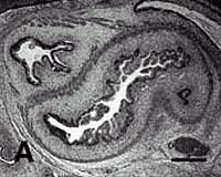
Cambridge MA (SPX) Jun 14, 2004 An MIT team has developed new technology that could jump-start scientists' ability to create specific cell types from human embryonic stem cells, a feat with implications for developing replacement organs and a variety of other tissue engineering applications. The scientists have already identified a simple method for producing substantially pure populations of epithelial-like cells from human embryonic stem cells. Epithelial cells could be useful in making synthetic skin. Human embryonic stem cells (hES) have the potential to differentiate into a variety of specialized cells, but coaxing them to do so is difficult. Several factors are known to influence their behavior. One of them is the material the cells grow upon outside the body, which is the focus of the current work. "Until now there has been no quick, easy way to assess how a given material will affect cell behavior," said Robert Langer, the Germeshausen Professor of Chemical and Biomedical Engineering. Langer is the senior author of a paper on the work that will appear in the June 13 online issue of Nature Biotechnology. The new technique is not only fast; it also allows scientists to test hundreds to thousands of different materials at the same time. The trick? "We miniaturize the process," said Daniel G. Anderson, first author of the paper and a research associate in the Department of Chemical Engineering. Anderson and Langer are coauthors with Shulamit Levenberg, also a chemical engineering research associate. The team developed robotic technology to deposit more than 1,700 spots of biomaterial (roughly 500 different materials in triplicate) on a glass slide measuring only 25 millimeters wide by 75 long. Twenty such slides, or microarrays, can be made in a single day. Exposure to ultraviolet light polymerizes the biomaterials, making each spot rigid and thus making the microarray ready for "seeding" with hES or other cells. (In the current work, the team seeded some arrays with hES and some with embryonic muscle cells.) Each seeded microarray can then be placed in a different solution, including such things as growth factors, to incubate. "We can simultaneously process several microarrays under a variety of conditions," Anderson said. Another plus: the microarrays work with a minimal number of cells, growth factors and other media. "That's especially important for human embryonic stem cells because the cells are hard to grow, and the media necessary for their growth are expensive," Anderson said. Many of the media related to testing the cells, such as antibodies, are also expensive. In the current work, the scientists used an initial screening to find especially promising biomaterials for the differentiation of hES into epithelial cells. Additional experiments identified "a host of unexpected materials effects that offer new levels of control over hES cell behavior," the team writes, demonstrating the power of quick, easy screenings. Community Email This Article Comment On This Article Related Links MIT SpaceDaily Search SpaceDaily Subscribe To SpaceDaily Express Space Medicine Technology and Systems
 Richland WA (SPX) Jun 22, 2005
A new computational tool developed at the Department of Energy's Pacific Northwest National Laboratory is speeding up our understanding of the machinery of life - bringing us one step closer to curing diseases, finding safer ways to clean the environment and protecting the country against biological threats. |
|
| The content herein, unless otherwise known to be public domain, are Copyright 1995-2006 - SpaceDaily.AFP and UPI Wire Stories are copyright Agence France-Presse and United Press International. ESA PortalReports are copyright European Space Agency. All NASA sourced material is public domain. Additionalcopyrights may apply in whole or part to other bona fide parties. Advertising does not imply endorsement,agreement or approval of any opinions, statements or information provided by SpaceDaily on any Web page published or hosted by SpaceDaily. Privacy Statement |