 |
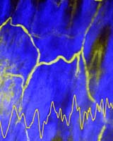
New York (SPX) Jun 02, 2004 Biomedical microscopic imaging deep inside living tissue with unprecedented clarity could become routine and widely available with the signing of technology-transfer and collaborative-research agreements on Friday (May 28, 2004) by Carl Zeiss Jena GmbH, a leading maker of microscopy instrumentation, and by CCTEC, the technology, enterprise and commercialization arm of Cornell University. The license for two-photon laser microscopy (also known as multiphoton microscopy, and protected by patents dating back to July 23, 1991) has been transferred from the British firm Bio-Rad Laboratories to Germany's Carl Zeiss. Both Bio-Rad and Carl Zeiss have been manufacturing confocal laser microscopes incorporating multiphoton technology. Additionally, Carl Zeiss has signed collaboration and development agreements with Cornell, in Ithaca, N.Y., and with multiphoton microscopy co-inventor Watt W. At Cornell, Webb, a biophysicist, is director of the National Institutes of Health-funded Developmental Resource for Biophysical Imaging and Opto-electronics (DRBIO) and is the S.B. Eckert Professor in Engineering. Co-inventor Winfied Denk is a director of Germany's Max-Planck-Institute for Medical Research in Biomedical Optics. Multiphoton microscopy produces high-resolution, three-dimensional images of tissues -- in the central nervous system, for example, or in pre-cancerous cells -- with minimal damage to living cells. The procedure begins when extremely short, intense pulses of laser light are directed at cells below the surface. The rapid-fire nature of multiphoton microscopy increases the probability that two or three photons will interact with individual biological molecules at the same time, combining their energies. The cumulative effect is the equivalent of delivering one photon with twice the energy (half the wavelength, in the case of two-photon excitation) or three times the energy (one-third the wavelength in three-photon excitation) to illuminate the smallest details. As a scanning laser microscope moves the focused beam of pulsed photons across a sample at a precise depth (plane of focus), cells above or below the plane are not affected. When repeated scans at different focal planes are "stacked" by computer processing, a brilliant, three-dimensional picture emerges. Initially developed at Cornell to enhance basic biological research, multiphoton microscopy is proving to be a marked improvement over existing biomedical-imaging techniques. Wherever it is conducted -- in imaging of still-living tissue outside the body(ex vivo) or endoscopic imaging practically anywhere in the body(in vivo) -- multiphoton microscopy depicts cells and cellular processes in vivid, microscopic detail across the third and fourth (time) dimensions. Ulrich Simon, head of microscopy at Carl Zeiss, said the Zeiss acquisition of Bio-Rad's Cell Science Division plus the collaboration with Cornell's DRBIO laboratory, and the German firm's experience in developing and manufacturing scientific instruments, will bring together the strongest forces in laser scanning microscopy to benefit biomedical science. "Today, Carl Zeiss stands for high-end laser scanning microscopes that are benchmark solutions for advanced applications in 3-D microscopy," Simon said. "Joining our forces will strengthen our relationship with Zeiss and Bio-Rad customers and will allow a considerable additional refinement of both companies' worldwide service and support network." Bio-Rad, a pioneer in developing confocal microscopy and multiphoton technology, has had a long-term collaborative partnership with Cornell, said Webb. "Now, working with Carl Zeiss should provide access to a new source of optical and instrumental expertise. Our objective is to further the technology and enable demanding applications of our powerful multiphoton microscopy technology." Webb said the biomedical research community is beginning to recognize the advantages of multiphoton microscopy "to address their research questions in the most natural context -- deep in the tissue or even directly in the living organisms. Our research is focused on developing the potential of the technology, as well as on applying it to solve 'impossible' biological problems," Webb said. "Apart from imaging deep in the tissue, multiphoton microscopy and nonlinear optics have several advantages for breakthrough applications in biomedical research." One such application combines multiphoton microscopy with fluorescence correlation spectroscopy (invented by Webb and by Elliot Elson and Doug Magde decades ago) to measure the dynamics of biomolecular processes of sparse biomolecules in living cells, Webb said. "Another surprisingly useful feature [of multiphoton microscopy] is imaging of the intrinsic fluorescence of biomolecules deep in living plant and in animals, where it can diagnose many disease states." Related World Wide Web sites: The following sites provide additional information on this news release. Some might not be part of the Cornell University community, and Cornell has no control over their content or availability. Community Email This Article Comment On This Article Related Links Carl Zeiss International DRBIO SpaceDaily Search SpaceDaily Subscribe To SpaceDaily Express Space Technology News - Applications and Research
 Baltimore MD (SPX) Jan 12, 2006
A team comprised of three leading US aerospace and defense contractors has demonstrated an innovative technological use of active electronically scanned array (AESA) radars for high-bandwidth communications. |
|
| The content herein, unless otherwise known to be public domain, are Copyright 1995-2006 - SpaceDaily.AFP and UPI Wire Stories are copyright Agence France-Presse and United Press International. ESA PortalReports are copyright European Space Agency. All NASA sourced material is public domain. Additionalcopyrights may apply in whole or part to other bona fide parties. Advertising does not imply endorsement,agreement or approval of any opinions, statements or information provided by SpaceDaily on any Web page published or hosted by SpaceDaily. Privacy Statement |